Stroke imaging analysis
Advanced imaging (via MRI, conventional CT, and flat-panel CT) provides a crucial window into the status of brain tissue during an acute stroke, allowing the identification of tissue with irreversible ischemic damage, and of salvageable tissue with compromised blood supply (the so-called ‘penumbra’). Current guidelines recommend the use of advanced imaging for patient selection where patient present between 6 and 24 hours after stroke: however, current imaging concepts for treatment selection are based on fixed thresholds which cannot take into account substantial variations in centre, scanner and sequence.
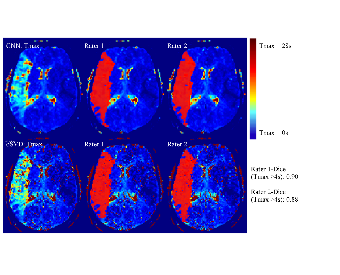
Leveraging modern techniques in machine learning and computer vision, we develop methods for forecasting the outcome of successful intervention, versus no intervention, based on a patient’s imaging and risk factors. These methods include harmonization of stroke imaging between centres and devices, accurate identification of tissue at risk and persistent infarction, fast generation of perfusion maps using neural networks, and lesion-load mapping to identify eloquent areas at risk of ischemic damage.